
Numerical Model
Numerical technique:
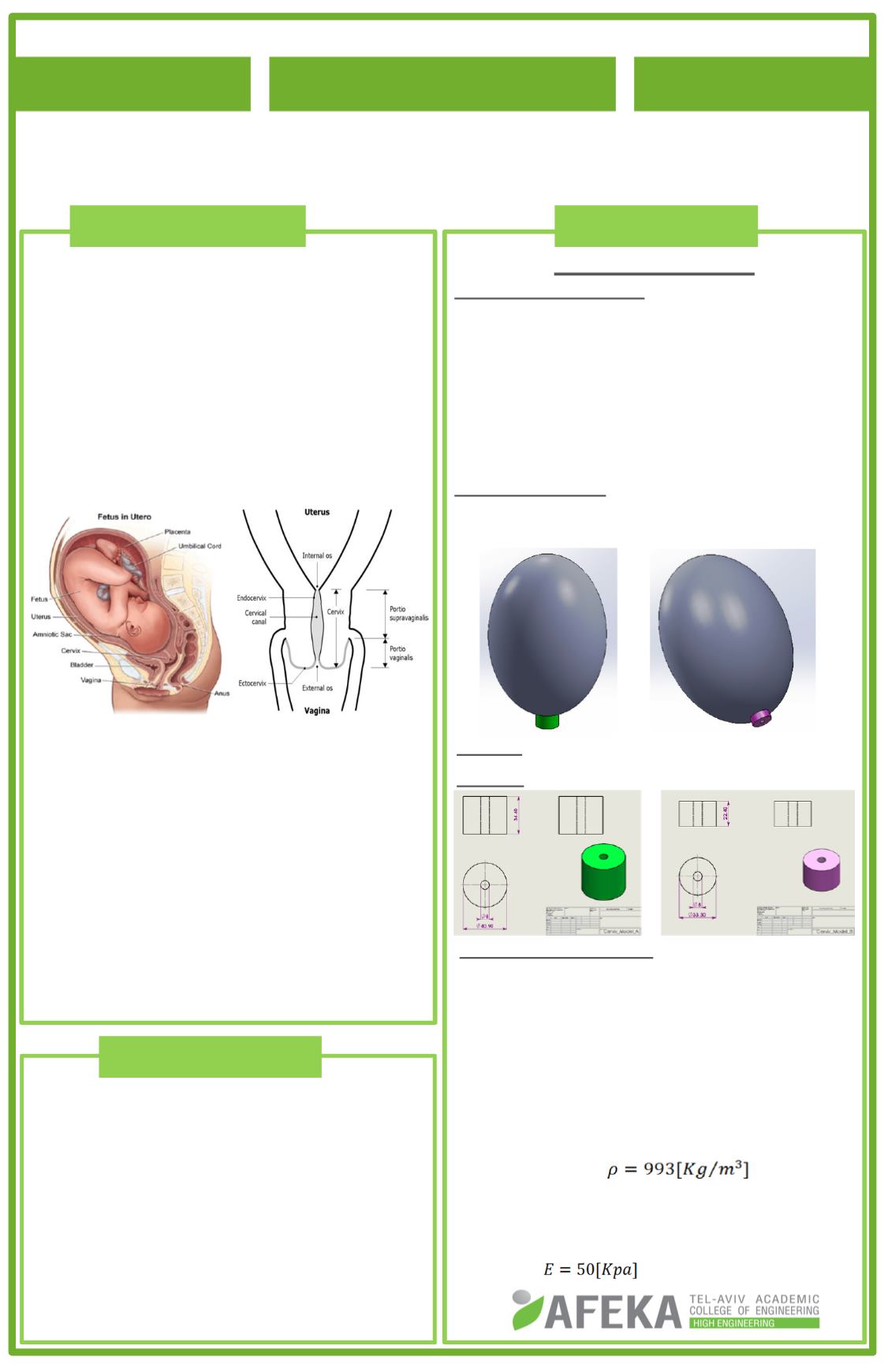
Geometrical model assembly:
SolidWorks 3D CAD.
Numerical Simulation package:
ANSYS 14.5, implements the FEM.
Elements Type: Tetrahedral with 4 nodes
Mesh: 11,600 elements for model A,
15,700 elements for model B.
Model Geometry - compatible with the 37
th
week of gestation: uterus is 290mm long,
210mm wide and 10mm thick.
Case A: Maximal cervical dimensions.
Case B: Minimum cervical dimensions.
Boundary Conditions:
The uterus is fixed at its upper part.
The cervix is fixed at the bottom, around
its exterior diameter.
To simulate the support of pelvic
ligaments, a thin disk was added
surrounding the top of the cervix, and was
fixed around its outer scope.
The entire uterine cavity is filled with
amniotic fluid ( ), which
puts hydrostatic pressure on the model.
The models were applied with the normal
elasticity modulus of uterine and cervical
tissue ( ).
Student name:
Meital Bistri
Department: Medical Engineering
Advisor name: Dr.
Zoya Gordon
Analysis of the stresses evolving in
cervix during pregnancy
2015
---
1
The human uterine cervix maintains the
uterus closed during gestation to ensure
a normal development of the fetus.
As pregnancy progresses, uterine volume
and pressure increase and the cervix has
to withstand larger forces, including
dynamic fluctuations.
The competent cervix stays firm and its
canal is closed.
Cervical incompetence is the inability of
the cervix to conserve the developing
pregnancy, which usually leads to
preterm delivery.
Early detection of an incompetent cervix
is important in helping to prevent preterm
labor. However, the diagnosis of cervical
incompetence is very difficult to make.
The project studies the behavior of the
cervix under the influence of the stresses
exerted on it during pregnancy.
Analyzing the distribution of the
stresses and strain evolving in cervix
during pregnancy.
To investigate the correlation between
the geometry of the cervix, and the
stresses, leading to its deformation, while
taking into consideration the mechanical
properties of the cervical tissue.
A
B
2. Objectives
1. Background
3. Methods


